What to expect
About Your Visit
We aim to make your first visit and every visit thereafter as pleasant as possible and what is better than knowing what to expect? When you visit us for the first time we will ask you to fill in a form providing us with your personal information as well as that of your medical aid. When you see the doctor he will take your medical history and do a clinical evaluation. It is helpful if you bring along any medication you are currently taking and any cardiac tests that your family physician has conducted. If the doctor suspects any abnormalities he will ask for a few tests to be performed by the clinical technologist. For more information on these tests please refer to Our Services.
Our services
Resting ECG (electrocardiogram): A 12 lead ECG is a recording of the heart’s electrical activity while the patient lies down. The recording is used to show the conduction through the heart which may be affected by underlying heart diseases. It can also show structural abnormalities such as chamber dilatation and hypertrophy, evidence of a previous heart attack and ischeamia as well as abnormal rhythms. The test will require you to remove the clothes off your chest. Should you have a lot of chest hair, areas of your chest may have to be shaved. Your skin will then be cleaned with an alcohol swab to remove any body lotion, sweat or powder. Small stickers called electrodes will then be placed in specific areas on your skin and cables will then connect to these electrodes to show the ECG on the monitor. This test usually takes about 10 minutes.
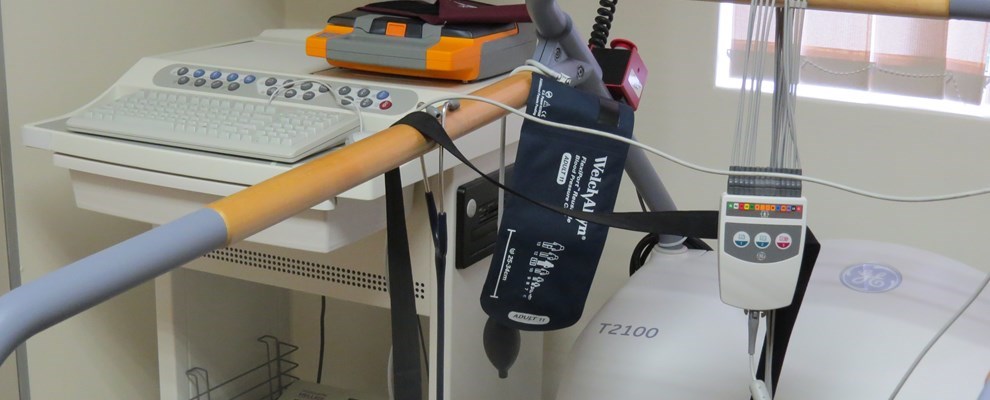
Stress/Exercise ECG: This is a 12 lead ECG recording of the heart’s electrical activity while you exercise. We start by obtaining an ECG while you rest to record a baseline ECG, heart rate, and blood pressure. You will then be asked to walk on a treadmill which progressively becomes faster and you may after a while even have to take a slight jog. Throughout the test the ECG is closely monitored for any abnormalities. The purpose of the test is to see whether the heart receives enough blood particularly during exercise. Preparation for this test is the same as for the resting ECG, the only difference is that you will be asked to walk and possibly run on a treadmill. You do not need to bring exercise clothes or tekkies with, but if you prefer to you are welcome to do so.
Holter/Event Monitor: This is a 24 to 48hours ECG monitor that makes a recording of the heart’s electrical activity. It is specifically used to monitor your heart rate and rhythm while you go on with your everyday routine. The monitor is battery operated. The purpose of the test is to document abnormal heart rates and rhythms. The test will require you to remove the clothes off your chest. Should you have a lot of chest hair, areas of your chest will have to be shaved. A few electrodes attached to cables will be placed onto your chest and attached to a small monitor. You will be asked to avoid swimming or bathing to prevent the system from getting wet. You will also be asked to keep a diary of your activities and associated symptoms.
Echocardiogram (ultrasound of the heart): This test uses sound waves to obtain a moving image of the internal structures of the heart. It can be used to measure the cavity size of the different chambers, see structural abnormalities, diagnose abnormalities of the heart valves and determine how well the heart is contracting and if the muscle has been damaged by a previous heart attack. The test will require you to remove the clothes off your chest, so that the technologist can place a probe with gel on various areas of your chest to obtain the images. You will feel some pressure but it is not painful.
Coronary Angiogram: We may recommend that you have a coronary angiogram if you have symptoms of coronary artery disease, such as chest pain (angina), pain in your jaw, neck or arm that can’t be explained by other tests or new/increasing chest pain (unstable angina). A coronary angiogram is a procedure that uses X-ray imaging to see your heart’s blood vessels. During a coronary angiogram, a type of dye that’s visible by an X-ray machine is injected into the blood vessels of your heart. The X-ray machine rapidly takes a series of images, showing a detailed look at the inside of your blood vessels. If necessary, your doctor can perform procedures such as angioplasty or stenting during your coronary angiogram.
For this procedure you will have to be admitted to the hospital and go to the cardiac catheterization laboratory. Here you will lie on a bed covered with sterile drapes. The doctor together with the nursing staff and technologist will do the procedure. There are two points of possible access into the body's blood vessels: the groin or the wrist. The doctor will administer a local anesthetic to numb the area, you will still feel him working in the area, but shouldn’t experience any pain. He will then use a needle to gain access to the arterial system (groin: femoral artery / wrist: radial artery). A small plastic tube will then be inserted into the artery and advanced to the heart where it will be used to administer the dye used to view the different arteries of the heart. Without any complications patients should be able to go home the same or the following day.
Coronary Angioplasy and Intervention: If a coronary angiogram is performed and it is found that there is a significant narrowing in one or more of the heart's blood vessels the doctor will decide which treatment plan is most suitable for the patient: should the patient go for a Coronary Artery Bypass operation or can they be helped by percutaneous coronary intervention (PCI).
PCI usually refers to 2 procedures:
1. Angioplasty: Often also called PTCA ( Percutaneous Transluminal Coronary Angioplasty). This will involve using a small balloon determined by the size of your vessels and the size needed to stretch open the narrowed segment in the artery upon inflation. The balloon is advanced to the narrowing and then inflated to press the plaque against the artery walls and open it to flow again.
2. Stenting: This will involve using a small spring like stent to stretch open the vessel and keep the plaque against the artery wall. The stent is compressed over a balloon, and like with angioplasty will have to be advanced to the narrowed area. It is then inflated and the stent is deployed in the vessel. The balloon can then be deflated and removed while the stent stays behind.
Device Implantation
- A pacemaker is a small device that is used to help treat abnormally slow heart rate/rhythms. This device uses electrical impulses to prompt the heart to beat at a normal rate. - Implantable cardiac defibrillators are used to monitor and treat abnormally fast heart rates/rhythms by delivering a shock to the heart to regulate the rhythm.
The procedure of implanting a pacemaker or implantable cardiac defibrillator usually takes about 45 minutes. It does not require open-heart surgery, and most people can be discharged within 24 hours. The procedure is performed under local anesthesia. A small incision, approximately 5cm long will be made below the collar bone on the left or right side of the chest. One, two or three leads (thin insulated wires) will be guided through a vein into the heart (the amount of leads depends on the type of pacemaker that you will get as they are implanted for different reasons). The doctor will then connect the lead(s) to your pacemaker and insert the device beneath your skin. The incision will be closed with dissolvable stitches. The doctor will test the pacemaker to ensure it is working properly to meet your medical needs.
For this procedure you will have to be admitted to the hospital and go to the cardiac catheterization laboratory. Here you will lie on a bed covered with sterile drapes. The doctor together with the nursing staff and technologist will do the procedure. You will be awake throughout the procedure as only local anesthesia is used. You will still feel the doctor working in the shoulder area, but it shouldn’t be painful. After the procedure you will stay in the hospital overnight and go home the following day with instructions on caring for your incision. You will be asked to limit how much you move the arm that is closest to your implant site for about 10 to 14 days after the surgery. After the implant, there may be a slight bulge visible under the skin where the device is located.
Device Testing and Programming
Pacemakers and Implantable Cardiac Defibrillators need to be tested frequently. 6 to 12 weeks after implantation you will come back for your first check-up. During this visit the area where the pacemaker was implanted will be checked for infection, you will have to go for a chest X-ray to check the pacemaker and lead(s) position and the pacemaker will be tested and adjusted if necessary. Depending on the type of device that you received you will be followed-up every 3, 6 or 12 months. These appointments are very important as your heart can become dependent on the pacemaker and the pacemaker battery only lasts a few years. Pacemakers can last an average of 5 – 10 years, whereas implantable cardiac defibrillators can last 3 - 5 years.
For pacemakers or implantable cardiac defibrillators testing and programming are done in the doctor’s rooms. You will lie down while the technologist connects you to an ECG and place a specific magnet called an analyzer head onto your pacemaker. The device will then be interrogated automatically. Some patients can feel their heart rate increasing during this test. The doctor or technologist will then perform a few tests on your device to optimize it for your specific needs.